Skeletal System
A. Body wall:
Turbellarians lack a cuticle, instead they have an epidermis that is one-celled thick (monolayered) and the cells are covered with small, hair-like structures known as cilia. (Fig.1-A). Beneath this is the basal lamina (Fig. 1-B), and together with the intracellular fibres, they acts as a skeletal support for the animal. The epidermal layer is also filled with pigment granules (Fig. 1-C)which accounts for their bright colour and patterns.
B. Parenchyma (connective tissue)
Figure 1. Cross section of parenchyma tissue in polyclads
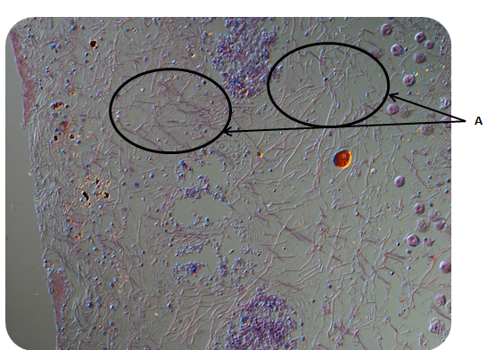 Figure 2. Frontal section of cuticle margin shows parenchyma tissue (A) that fills up the body cavity
 Large turbellarians (macroturbellarians) such as P. bedfordi have connective-tissue between the body wall muscles and gut called the parenchyma. Within this parenchyma , there are several types of parenchymal cells.
Some examples of these specialized cells are:
(i) Epidermal replacement cells (Fig.1-C)
When surface layer cells are damaged, these cells move up from the parenchyma and replace the destroyed cells at the surface.
(ii) Neoblasts
They have a population of totipotent cells known as neoblasts (Fig. 1-D). These are involved in wound healing and regeneration. They also give rise to the aforementioned epidermal replacement cells.
(iii) Fixed parenchymal cells (Fig.1E)
These are large, branched cells which make gap junctions with other parenchymal cells, gastrodermal and epidermal cells. They form a network of channels for low-resistance transport of cells.
(iv) Pigment cells and chromatophores (Fig1-F)
Pigment cells (Fig. 1) give the flatworm its distinctive colour on the body surface. Chromatophores are pigment-containing cells controlled by the nervous system and allows the flatworm to lighten or darken its body colour by concentrating or diffusing the pigment cells.(Ruppert et al., 2004)
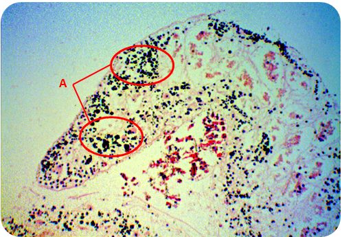
Figure 1. Frontal section of P. bedfordi pseudotentacles showing clusters of black pigment granules
(v)Glandular epidermis
Several gland cell types are embedded within the epidermis, the most common of which are the rod-shaped rhabdites (Figs. 2 & 3), which are secreted by epidermal gland cells. These gland cells secrete mucus and other adhesives to enable the flatworm to attach and glide on surfaces.
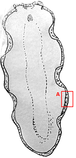
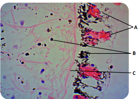
Figure2. Frontal section of cuticle margin from P. bedfordi showing submerged rhabdite gland (A), black pigment granules (B) and basal lamina (C).
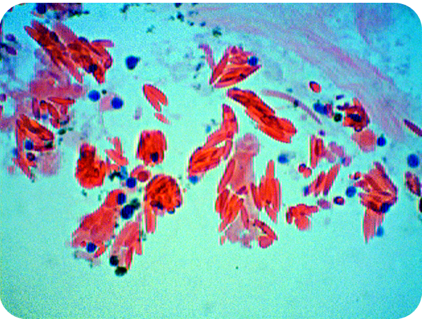
Figure 3. Frontal section of cuticle margin showing a close-up view of rod-shaped rhabdites secreted from rhabdite glands.